
Benjamin Plackett in Nature:
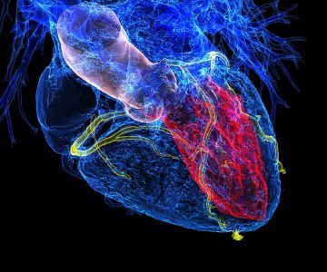 Current methods for predicting heart attacks are woefully outdated, says Cheerag Shirodaria, a cardiologist at the Oxford University Hospitals NHS Foundation Trust, UK. “We basically look at a patient’s age, obesity and whether they have diabetes. The most sophisticated we get is measuring their cholesterol,” he says. Heart attacks are often caused when fatty accumulations inside arteries, known as plaques, result in the narrowing of blood vessels. These plaques then rupture and form blood clots. Age, being overweight or obese and having diabetes are all risk factors, and people in at-risk groups are closely monitored. “But 50% of heart attacks are in people without narrowing, and they’re being missed by current tests,” he says. A desire to drag prognosis into the modern era is what pushed Shirodaria and a group of like-minded cardiologists to set up Caristo Diagnostics. Rather than blood-vessel narrowing, the team focuses on another driver of plaque ruptures — vascular inflammation. The company uses algorithms to analyse information that until now has been hidden in the noise of computed tomography (CT) scans. This enables it to assess inflammation and calculate the likelihood that a person will have a heart attack in a given time period.
…Fat tissue bordering blood vessels, known as perivascular fat, had long been thought to be a cause of inflamed arteries. “There was this perception that perivascular fat was the bad guy,” says Antoniades. That’s why his research started out as a hunt for any potential chemicals produced and sent by the fat to the blood vessels — the working theory used to be that such chemicals could have been behind the problem. “But the results always showed the opposite,” he explains. The signals were instead going the other way. Antoniades showed that when an artery is inflamed, it sends out SOS signals that are then picked up in the surrounding fat1. In response, the fat undergoes lipolysis — the lipids break down and the water content in the fat cells increases (see ‘Sensing a heart at risk’). That effectively means that diminished perivascular fat is an indicator of inflammation, and therefore also of an increased risk of heart attack. Other scientists have validated the link between perivascular fat abnormalities in CT scans and coronary inflammation2.
More here.