Rajagoalan and Long in Nature:
 Scientists have long puzzled over the ‘immunological paradox’ of pregnancy1: how does the mother tolerate the fetus — a foreign entity that carries some of the father’s DNA? In a paper in Nature, Vento-Tormo et al.2 investigate this enigma. The authors performed single-cell RNA sequencing (scRNAseq) of cells isolated from the placenta and the decidua (the lining of the pregnant uterus), and from matching maternal blood for comparison. They identified an array of cell types unique to this maternal–fetal interface, and inferred the existence of a large network of potential interactions between them that would favour immunological tolerance and nurture the growth of the fetus. The authors’ molecular atlas provides an impressive resource for future studies of pregnancy and its complications.
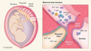
The early embryo develops into a structure called the blastocyst, which implants in the lining of the uterus. Implantation triggers the development of the placenta from fetal membranes. The placenta nourishes the fetus through the umbilical cord3. Abnormal placental development can lead to several complications of pregnancy, including pre-eclampsia, fetal growth restriction and stillbirth. A better understanding of human placental development is sorely needed, but there is no good animal model for this process — it has to be studied in women. Vento-Tormo et al. collected placental, decidual and blood samples from pregnancies that had been electively terminated at between 6 and 14 weeks of gestation. The authors’ scRNAseq analysis enabled them to distinguish between cells of maternal and fetal origin, because the latter include RNA sequences that are absent in the mother. This clearly revealed that cells from the fetus had migrated into the maternal decidua (Fig. 1), and that a small subset of maternal immune cells called macrophages were located in the placenta.
More here.
