
Josie Glausiusz in Nature:
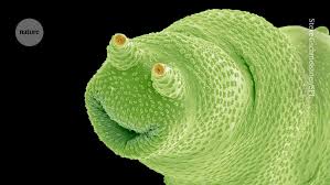 Cheese fungus, head lice, human sperm, a bee eye, a microplastic bobble: scientific photographer Steve Gschmeissner has imaged them all under the probing lens of a scanning electron microscope (SEM). In his colourized electron micrographs, faecal bacteria resemble thin spaghetti, silica-walled diatoms look like cubes of breakfast cereal and a segmented tardigrade resembles a curled-up, tubby piglet. Gschmeissner, who has been imaging microbes, cancer cells and invertebrates for about 50 years, has crafted an extraordinary array of more than 10,000 SEM images, some of which have been featured in Nature. He spoke to Nature about the importance of scientific images, looking at imploding cancer cells and the miniature world he found on a rotten raspberry.
What are some of the projects you’ve worked on recently?
For the past six years, I’ve been collaborating with Greg Towers, a molecular virologist at University College London, who supplies me with samples to photograph. We’ve looked at a variety of viruses, including SARS-CoV-2, which causes COVID-19. The latest work I’ve done with Towers is a project on cancer-cell death. It’s the sort of work I love doing: science that tells a story with images. It’s been one of my most enjoyable and successful recent projects, because there’s very little else out there that shows what happens to cancer cells during chemotherapy.
More here.